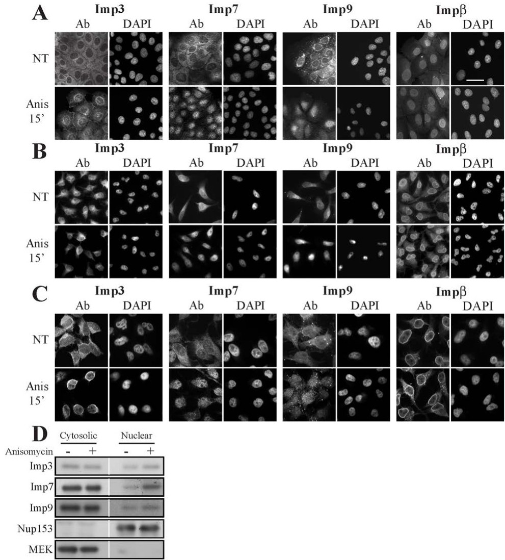
Fig. 7. The subcellular localization of Imps3, 7 and 9 in resting and stimulated cells. Fluorescent microscopy detects the nuclear shuttling of Imp3/7/9. HB2 (A) or HeLa (B, C) cells were grown on slides to 70% confluence, serum starved, and then either stimulated with anisomycin (Anis 0.5 μg/ml 15 min) or left untreated (NT). Cells were fixed and stained using the indicated Abs. The nuclei were detected using DAPI and slides were visualized using either a fluorescent microscope (A, B) or Zeiss Spinning Disk confocal microscope (C). The bar in the upper right panel of A is of 20 µM. (D) Subcellular fractionation confirms the nuclear translocation of Imps 3,7 and 9. The same samples of Fig. 1D were used here. Serum-starved HeLa cells were either stimulated (Anis, 0.5 µg/ml, 15 min) or left untreated then harvested. Subcellular fractions of cytosolic and nuclear were produced as described, and subjected to Western blot analysis with the indicated Abs.